Scoliosis
Reviewed by our Healthcare Team Member

Zihan Masood, MD
Share This Page
Scoliosis: Understanding, Diagnosing, and Treating Spinal Curvature in the 21st Century
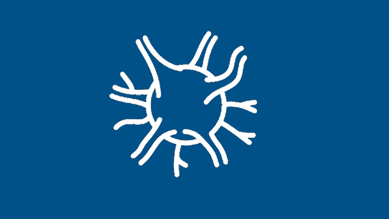
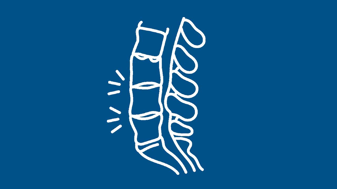
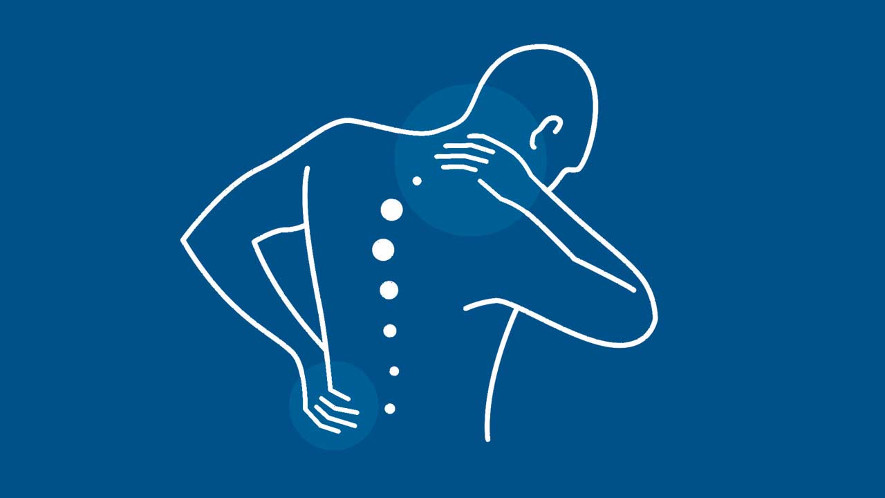
Scoliosis is a condition characterized by an abnormal curvature of the spine, which can be identified through imaging techniques such as X-rays or observed through visible postural imbalances, including thoracic rib humps or winging scapula. Recent research has revealed that patients with idiopathic scoliosis often exhibit asymmetry in the brain (cortex and cerebellum), mirroring the asymmetry in their spine. Furthermore, individuals with idiopathic scoliosis are more likely to experience neuropsychiatric conditions such as depression and schizophrenia. Understanding the connection between scoliosis and brain function provides valuable insights into treatment approaches.
Idiopathic Scoliosis: Genetic Onset, Non-Genetic Progression
Idiopathic scoliosis typically manifests during puberty and is believed to have a polygenetic basis. However, the progression of scoliosis is not genetic and is influenced by modifiable risk factors. While scoliosis is visually defined by the rotary deformity of the spine, the underlying causes involve brain asymmetry and metabolic imbalances. Addressing these factors early can prevent progression and improve patient outcomes.
A Shift in Treatment Paradigm
Traditionally, mild scoliosis has been managed through observation and monitoring without intervention. However, this approach often allows progressive curvatures to worsen, leading to irreversible bone deformities and the need for bracing or surgical correction. Untreated scoliosis can result in chronic pain, brain atrophy, postural asymmetry, and reduced quality of life. Immediate intervention following diagnosis is recommended to address functional deficits and mitigate risk factors for progression, ultimately improving patient outcomes.
The Brain-Spine Connection in Scoliosis
The spine, composed of 33 vertebrae, functions as a flexible structure supported by muscles that communicate positional information to the brain. In scoliosis, the spine rotates and shifts laterally, forming an "S" or "C" shape visible on imaging. This abnormal curvature can affect the thoracic, lumbar, or both regions, leading to muscle strain, disrupted alignment, and potential organ dysfunction. In adolescents, hormonal changes during puberty drive asymmetry, while in adults, brain asymmetry and sensory processing abnormalities contribute to motor signaling issues, further exacerbating spinal deformities. Effective treatment plans should address these underlying neurological and metabolic factors, rather than focusing solely on the spinal deformity.
Types and Causes of Scoliosis
- Idiopathic Scoliosis: The most common type, typically developing during adolescence due to asymmetric growth. Onset is linked to genetic predisposition, while progression is influenced by epigenetic factors.
- Congenital Scoliosis: Present at birth due to vertebral malformations during development, often associated with other organ or skeletal abnormalities.
- Neuromuscular Scoliosis: Results from neurological or muscular conditions such as cerebral palsy, muscular dystrophy, or spina bifida, leading to muscle imbalance and poor spinal control.
- Degenerative (Adult-Onset) Scoliosis: Caused by brain-based imbalances, movement asymmetry, and inflammation, often associated with age-related spinal degeneration.
- Secondary or Functional Scoliosis: Arises from external factors such as leg length discrepancies, muscle spasms, or posture imbalances, and typically resolves when the underlying issue is addressed.
Risk Factors
- Children and Adolescents: Rapid growth during ages 10–15 increases risk.
- Females: Higher likelihood of developing severe curves requiring treatment.
- Older Adults: Age-related degeneration can lead to progression.
- Metabolic Factors: Low bone density, increased inflammatory markers, and vitamin D deficiency are associated with scoliosis progression.
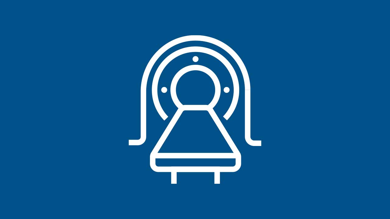
Diagnosing Scoliosis
A comprehensive diagnostic approach is essential to address both the curvature and the underlying neurological and metabolic imbalances driving progression. This includes:
- Physical Examination: Assessing spinal alignment, posture, and flexibility using advanced imaging techniques such as 3D rasterstereography.
- Neurological Testing: Vestibular and oculomotor assessments, computerized dynamic posturography, and EEG, EMG, and VEMP testing to identify brain asymmetry and sensory dysfunction.
- Metabolic Testing: Blood work to evaluate vitamin D levels, inflammatory markers, and bone density through DEXA scans.
Treatment Options Based on Curvature Severity and Age
- Mild Curvature (<25°): Early intervention is recommended to address neurological and metabolic imbalances. Flexible bracing may be considered.
- Moderate Curvature (25°–40°): Rigid bracing is typically required for adolescents, while adults may benefit from flexible bracing and other interventions.
- Severe Curvature (>40°–50°): Surgical intervention may be necessary, along with pain management and other treatments.
Non-Surgical Treatment Options
- Early Intervention: Addressing risk factors immediately upon diagnosis is critical to prevent progression. Lifestyle changes and targeted therapies should be implemented without delay.
- Bracing:
- Rigid Bracing: Effective for adolescents with curves between 25° and 45° to prevent progression during growth.
- Flexible Bracing: Suitable for adults and adolescents with smaller curvatures to reduce abnormal movement patterns.
- Custom Braces: Options include CMP overcorrection braces, nighttime bending braces, and Spinecor flexible braces.
- Rigid Bracing: Effective for adolescents with curves between 25° and 45° to prevent progression during growth.
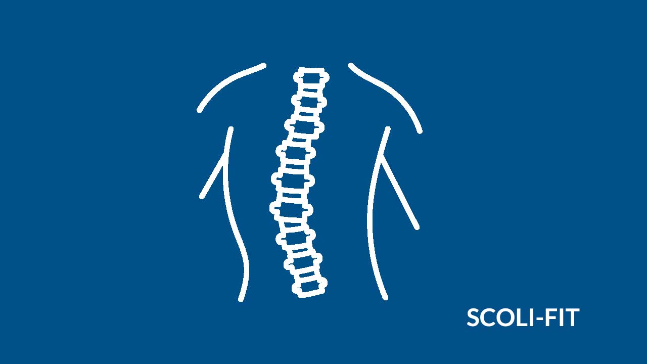
- Physical Therapy and Exercise: Focused on improving posture, spinal alignment, stability, and balance while reducing pain and fatigue. Techniques include the Scoli-Fit Schroth Method, core stabilization, and manual therapy.
- Pain Management: For adults with degenerative scoliosis, NSAIDs, muscle relaxants, and epidural steroid injections may alleviate discomfort.
Surgical Treatment Options
Surgery is typically reserved for severe cases with curves exceeding 45°–50°, significant pain, nerve compression, or impaired organ function. Options include:
- Spinal Fusion: Fusing curved vertebrae with rods, screws, and bone grafts for alignment and stabilization.
- Minimally Invasive Techniques: Robotic-assisted navigation, expandable rod systems for growing children, and vertebral body tethering (VBT) for select adolescents.
- Postoperative Recovery: Includes hospital stays, gradual return to activities, and physical therapy for optimal recovery.
Rehabilitation and Long-Term Care
Post-surgical physical therapy, ongoing exercise programs, and regular follow-ups are essential for maintaining alignment, promoting mobility, and preventing complications.
Living with Scoliosis
With early detection and comprehensive care, individuals with scoliosis can lead active and fulfilling lives. A combination of observation, bracing, physical therapy, and advanced surgical techniques ensures improved quality of life and minimal long-term limitations.
Multi-Disciplinary Approach
Our New York City-based practice offers integrated care for scoliosis patients, combining expertise from neurosurgeons, chiropractic neurologists, physical therapists, orthopedic spine surgeons, physiatrists, and pain specialists. We provide personalized treatment plans to restore alignment, alleviate pain, and enhance function, helping patients achieve optimal spine health.
Additional Resources
- National Institute of Arthritis and Musculoskeletal and Skin Diseases (NIAMS): Scoliosis
- American Association of Neurological Surgeons : Scoliosis Overview
Expert Care For This Condition Provided By

Alexandros Zouzias, MD

Marc Lamantia, DC

George Resnick, DC
Zihan Masood, MD